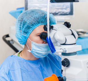
 At Family Eye Care & Surgery, our physician is committed to providing you with the highest quality eye care. Her surgical specialties include the following procedures:
- Cataract Surgery
- Corneal Surgery and Transplantation
- Eyelid Surgery
- Repair of Traumatic Pupil and Lens Damage
- Pterygium Excision with Ocular Surface Reconstruction
Cataract Surgery
Watch Dr. Herz discuss cataracts on the Hope Channel Network, the risk factors for getting cataracts, when to have them treated, and what to expect before and after cataract surgery.
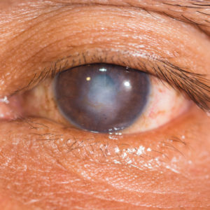
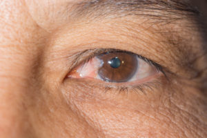
As we age, our eye’s natural lens begins to cloud resulting in blurry vision. This clouding of the lens is called a cataract. Once the cataract impairs your vision to the point it is interfering with your life, it is time for cataract surgery. This involves removing your natural, cloudy lens, and replacing it with a synthetic lens called an intraocular lens or “IOL.” Patients who receive cataract surgery can largely reduce their need for glasses or contacts depending on the IOL they choose. Find out if you can benefit from cataract eye surgery and intraocular lenses by asking our surgeons about the following choices:
- Monofocal IOL – now with UV protection and aspheric correction to improve night vision.
- Multifocal plus Toric IOL – the new “trifocal” lens that enables distance, computer and reading vision.
PanOptix Trifocal IOL – Only FDA-Approved Trifocal Lens - Toric IOL – fashioned as the monofocal IOL plus correction for astigmatism.
Download the AcrySof® IQ Toric Patient Education Brochure
Read more about cataracts in Focus Magazine: A journey to clearer vision (PDF)
Corneal Surgery and Transplantation
Our surgeon is a fellowship-trained cornea specialist and provides a wide range of medical and surgical treatment for corneal disorders including the following:
- Fuch’s Corneal Dystrophy
- Bullous Keratopathy
- Corneal Edema
- Keratoconus
- Corneal Ulcers
- Dry Eyes
She is experienced in corneal transplantation including DSAEK (Decemet’s Stripping Automated Endothelial Keratoplasty) as well as DMEK (Decemet’s Membrane Endothelial Keratoplasty). These procedures enable most patients with Fuch’s dystrophy, bullous keratopathy, and some cases of corneal edema to undergo a partial-layer corneal transplant that can shorten visual recovery by 6-12 months over traditional methods. Find out if you can benefit from one of these innovative techniques by calling for an appointment today.
Eyelid Surgery
- Blepharoplasty – Removal of excess eyelid skin that allows increased opening of the eye for greater peripheral vision and ease when reading.
Repair of Traumatic Pupil and Lens Damage
- Pupillary Cerclage – Somtimes cataract surgery or trauma to the eye can tear the iris and cause the pupil to lose its natural shape. This can cause great discomfort and decreased vision from bright sunshine and glare from headlights while driving at night. A pupillary cerclage repair can often restore the edges of the pupil back to a more normal shape during most lighting conditions.
- Traumatic Lens Damage – Severe ocular trauma can cause a cataract, often dislocating the natural lens of the eye. Our highly trained surgeon can remove the damaged lens and replace it with an intraocular lens that can be sewn into position if the natural support has been damaged.
Pterygium Excision with Ocular Surface Reconstruction
- Pterygium – degeneration of the conjunctiva due to sun and wind exposure that causes it to thicken, increase in blood vessels (causing the eye to often appear pink/red), and grow up onto the cornea.
- Surgical repair – If a pterygium is often inflamed and irritated, it will progressively grow towards the center of the pupil. When it gets too close to the pupil, it causes permanent loss of vision and surgical excision is required before it reaches this point. The surgery must include reconstruction of the area where the growth was, or it has a 50% chance of coming back. This is accomplished by harvesting an area of healthy conjunctiva from under the upper lid in the same eye and grafting it into the position where the growth was. This reduces the chance of recurrence to 5% and cosmetically improves the appearance of the eye.